Typically you suppose you perceive one thing, however you fail to clarify it. That is the time that you must look again from a special perspective and begin over. Whenever you dig in medical photos you will note completely different ideas to appear imprecise and non-intuitive, no less than to start with. You will notice individuals discussing DICOM and coordinate programs you have got by no means heard earlier than. Because of this, numerous misconceptions and confusions are born. If you’re on this place, or if you need to find out about AI in medical imaging this text is for you.
Again in 2017, once I utilized for my grasp’s diploma in biomedical engineering all people requested me why, as I used to be already obsessive about deep studying. Now, each multidisciplinary deep studying analysis challenge requires area data equivalent to medical imaging. Curiously, the funding within the AI Healthcare area is constantly growing. As an quantitative instance of first google search that one can discover out:
The marketplace for machine studying in diagnostic imaging will high 2 billion $ by 2023.
So, the rationale that I made a decision to jot down this text is to assist ML individuals dive into medical imaging.
In a earlier article, I talked a few widespread deep studying pipeline utilized to multi-modal magnetic resonance datasets. All of that in fact with our underneath growth open supply pytorch library known as medicalzoo-pytorch. Nevertheless, I did not dive into the particularities of the medical world an excessive amount of. In the long run, I used already processed information from an ML competitors (and never from a messy hospital), so any person else did the soiled work for me. This tutorial is partly primarily based within the nipy [1] and 3D Slicer.org [2] documentations for medical photos and Dicom information.
To dive deeper into how AI is utilized in Medication, you may’t go flawed with the AI for Medication on-line course, supplied by Coursera. If you wish to concentrate on medical picture evaluation with deep studying, I extremely advocate ranging from the Pytorch-based Udemy Course.
Nevertheless, I made a decision to adapt and revisit the ideas and make them extra acquainted to Machine and deep studying engineers. There are numerous assumptions that ML engineers do not know about. Different multi-disciplinary initiatives have this sort of terminology drawback. To this finish, I thought of it of nice worth to bridge this hole between medical imaging ideas and deep studying that nobody talks about, on this humble publish. At the least, I am going to strive my finest, one idea at a time!
Notation: medical picture tutorials typically name the MRI and CT exams as ‘mannequin’. For comfort, and to keep away from misconceptions we are going to use the world modality, all through this tutorial to check with any form of medical picture examination. The phrase ‘view’ and ‘aircraft’ are used interchangeably.
We’re all set to enter the medical imaging world!
The coordinate programs in medical imaging
A coordinate system is a technique for figuring out the placement of some extent. Within the medical world, there are three coordinate programs generally utilized in imaging functions: the world, anatomical, and the medical picture coordinate system.
Supply: 3D slicer documentation [2]
World coordinate system
The world coordinate system is a Cartesian coordinate system wherein a medical picture modality (e.g. an MRI scanner or CT) is positioned. Each medical modality has its personal coordinate system, however there is just one world coordinate system to outline the place and orientation of every modality.
Anatomical coordinate system
An important mannequin coordinate system for medical imaging strategies is the anatomical house (additionally known as the affected person coordinate system). This house consists of three planes to explain the usual anatomical place of a human:
-
the axial aircraft
-
the sagittal aircraft
-
the coronal aircraft
It’s price mentioning that this technique was invented for higher communication between docs and radiologists.
Tip: In an effort to perceive the anatomical planes, you must visualize a standing human taking a look at you.
Furthermore, on this system every part is related. Thus, the 3D place is outlined alongside the anatomical axes of anterior-posterior (front-back) and left-right and inferior-superior, as we are going to see. By this sense, all axes have their notation in a constructive route.
Axial aircraft
The axial aircraft is definitely once you place your perspective above the affected person and look down. Relying on the area of the 3D medical picture you’ll observe completely different anatomical buildings. For a 3D whole physique scan, should you had a control-bar over this 2D view you’ll begin from a 2D slice of the pinnacle, and by growing you’ll find yourself within the legs. Let’s virtually name this view the “drone aircraft” or “top-view”. Under you may see completely different slices of a mind MRI.

For anatomical consistency causes, a slice close to the pinnacle is known as superior in comparison with a slice nearer to the ft, which known as inferior.
Does it appear advanced? It’s, I do know! But it surely’s necessary on your psychological sanity if you wish to survive within the subject. Sadly, the opposite two planes assume completely different instructions for constructive 😢.
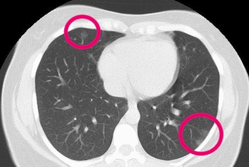
For completeness, and to current one thing a bit of bit more moderen under you see an axial CT of a affected person. Radiologists have highlighted irregularities that could be because of COVID. I’m simply resharing the picture right here for example the axial slice, trying from the highest to the affected person. The actual slice refers within the lungs.

The 30-year-old lady had patches that resemble frosted glass, according to COVID-19 pneumonia.(Equipped: Radiopaedia, Dr Bahman Rasuli)
Sagittal aircraft
Mainly, it is a facet view. As an alternative of trying from above the affected person, now we glance from the facet. The facet might be both proper or left. Which facet and route is the constructive one, depends upon the coordinate system! The positive factor is that from this view (aircraft) you may see the affected person’s ear! As you progress via this axis you see the projected tissues like lungs, bones.
A sagittal view of a mind MRI might be illustrated under:

Coronal aircraft
On this view, we traverse both by trying within the eyes (anterior aircraft) or by trying at the back of a affected person (posterior aircraft). I hope you get the thought by now.
In an effort to just be sure you will always remember what a coronal aircraft is, I discovered this terrible halloween gif:

The highlighted phrases in daring outline the completely different Anatomical coordinate programs, as illustrated under.
Tip: LPS is utilized by DICOM photos and by the ITK toolkit (simpleITK in python), whereas 3D Slicer and different medical software program use RAS.
Medical Picture coordinate system (Voxel house)
That is the half that comes extra intuitively for individuals with a pc imaginative and prescient background. You probably have any expertise with different 3D deep studying domains, I can guarantee you that that is the place that you’ll discover some rationality and related context, ultimately!
Medical modalities create 3D arrays/tensors of factors and cells which begin on the higher left nook, much like an RGB digital camera for 2D. The i axis will increase to the precise (width), the j axis to the underside (peak), and the okay axis backward (the third equally to the function maps of a conv internet).
Along with the depth worth of every voxel (i j okay), the origin (i.e. MRI) and spacing of the coordinates are saved within the meta-data of the medical picture (both dicom tags, header file of nifty photos, you title it.. ).
Voxel spacing is mainly the true measurement of the voxel that corresponds to a 3D area and the gap between two voxels.
That is vital if we wish for instance to measure the quantity of a most cancers tumor cell.
Observe that, it’s potential to resample to a much bigger voxel measurement to cut back the scale of the medical picture. This may be understood as a downsampling operation of a sign. Medical individuals will say that on this approach we preserve the sector of view the identical, however let’s merely say that it’s a form of downsampling method within the voxel house.
Observe: if the voxels are of the identical measurement in all 3 dimensions we name the picture isotropic, much like isotropic scaling in RGB photos.
Now that we see all these good coordinate theories, let’s see how we are able to manipulate and rework from one world to a different.
Transferring between worlds
In an effort to transfer from one world to a different, we’d like a magic key: it’s known as the well-known affine matrix. With this, we are able to transfer from one world to a different by way of a so-called affine transformation. However what’s an affine transformation?
Affine transformation
Earlier than affine transformation let’s make clear what a geometrical transformation is. A geometrical mapping/transformations is a approach to make clear that the voxel depth doesn’t change.
So, primarily based on the definition of Wikipedia for affine [7]: in geometry, an affine transformation is a geometric mapping of an affine house that preserves numerous properties such because it sends factors to factors, strains to strains, planes to planes. Moreover, it additionally preserves the ratio of the lengths of parallel strains. Nevertheless, an affine transformation doesn’t essentially protect angles between strains or distances between factors.
In math, to signify translation and rotation collectively we have to create a sq. affine matrix, which has another dimensionality than our house. Since we’re within the 3D house we’d like a 4D affine matrix in medical imaging. With the affine matrix, we are able to signify any linear geometrical transformation (translation, rotation), by a matrix multiplication, as illustrated under.
To this finish, we are able to go from the voxel house to world house coordinates of the imaging modality.
Observe: within the affine transformation, components listed by A signify translation and t-indexed components signify rotation.
Transferring from one modality to a different
We already noticed that the affine is the transformation from the voxel to world coordinates. The truth is, the affine was a fairly attention-grabbing property: the inverse of the affine offers the mapping from world to voxel. As a consequence, we are able to go from voxel house described by A of 1 medical picture to a different voxel house of one other modality B. On this approach, each medical photos “reside” in the identical voxel house.
Let’s see some compact code to carry out this operation:
import scipy
def transform_coordinate_space(modality_1, modality_2):
"""
Transfers coordinate house from modality_2 to modality_1
Enter photos are in nifty/nibabel format (.nii or .nii.gz)
"""
aff_t1 = modality_1.affine
aff_t2 = modality_2.affine
inv_af_2 = np.linalg.inv(aff_t2)
out_shape = modality_1.get_fdata().form
T = inv_af_2.dot(aff_t1)
transformed_img =
scipy.ndimage.affine_transform(modality_2.get_fdata(), T, output_shape=out_shape)
return transformed_img
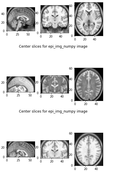
Let’s see what occurs if we apply this code in two completely different MRI photos:

Transferring between voxel areas
The primary 3 slices are from the referenced picture, the following 3 slices are from the picture that we need to rework, and the third one is the remodeled picture to the referenced world. They each reside in the identical affine world and we are able to visualize them facet by facet they usually even have the identical form.
Now that we briefly coated some coordinate system ideas, let’s focus on the DICOM nightmare!
All fashionable medical imaging modalities like X-Rays, Ultrasound, CT (computed tomography), and MRI (Magnetic Resonance Imaging) help DICOM and use it extensively. DICOM is to start with an Interface Definition. It’s success depends on the flexibility to combine medical modalities manufactured by completely different distributors. And sadly for the medical world, this was virtually not possible earlier than DICOM. For the file, integrating medical tools of various distributors was an enormous problem. Truly, that is the rationale that DICOM has naturally turn into the business commonplace.
DICOM is a singular format because it doesn’t solely retailer the medical picture information but in addition data-sets, that are made up of attributes. For our readers with a software program engineering background let’s merely say that it incorporates an enormous quantity of meta-data with the picture (and normally redundant). The meta-data include vital info that have to be stored throughout the file to make sure they’re by no means separated from one another.
Tip: To summarize, the core of DICOM is each a file format and a networking protocol.
-
DICOM File Format: All medical photos might be saved in DICOM format. Medical imaging modalities create DICOM information. The large adoption of DICOM information is justified as a result of they include extra than simply medical photos, as defined. Particularly, each DICOM file holds demographic affected person info (title, beginning date), in addition to necessary acquisition information (e.g., kind of modality used and its settings), and the context of the imaging research (i.e. radiotherapy collection, affected person historical past). As a machine studying engineer, that is probably the most vital idea that you could take care of Dicom information.
-
DICOM Community Protocol: Nevertheless, DICOM doesn’t solely outline the picture and data-sets (meta-data) but in addition the transport layer protocol, roughly like TCP or some other protocol. Your complete commonplace is made up of a number of associated however impartial sections. Nonetheless, all medical imaging functions which can be related to the hospital community use the DICOM protocol to trade info, which is principally DICOM photos. Furthermore, the DICOM community protocol is used to seek for imaging research within the archive and restore imaging research to the workstation with a purpose to show it. Lastly, every part advanced abstraction comes with a superb half: Since DICOM is a posh protocol it permits for a number of instructions equivalent to schedule procedures, report statuses.
Tip: In medical picture evaluation we’re largely all in favour of understanding our information so we are able to preprocess them to coach a deep neural community.
Any more, we are going to check with DICOM because the file format for simplicity.
DICOM and deep studying
In the event you work on a challenge with DICOM information, you in all probability need to examine a bit of little bit of the usual. However let’s begin with what we’d actually need to have to use our deep studying mannequin. So ideally, we’d simply need to do one thing like this for any modality:
import awesome_library
medical_img_volume_in_numpy = awesome_library.loadDicom(path)
This presently would not exist for DICOM due to the variety of the completely different modalities and the completely different particularities that exist within the Dicom photos. As an illustration, a computed tomography picture has completely different metadata (tags) than a magnetic resonance picture.
Nevertheless, should you handle to transform your information within the nifty format, there may be an superior library known as nibabel that does precisely what we wish.
Tip: there’s a magic library that reads nifty (*.nii) information – NOT DICOM- in Python known as nibabel that does what we needed within the first place.
We are going to element under on that.
Luckily, there are two superior python packages that may save us time: pyDICOM [5] and dcm2niix. With pyDICOM we are able to learn and manipulate Dicom information or folders. A few of chances are you’ll surprise why this operation isn’t trivial! Is it so tough to simply get a 3D NumPy array of values?
The reply is sure, just because normally Dicom information are normally organized as this: each single 2D picture slice is a special Dicom file. It’s common that the completely different exams from a number of modalities usually are not even in the identical folder. Actual-world medical information are messy!
dcm2niix
Alternatively, the superior software known as dcm2niix can convert a DICOM folder that incorporates a multi-sequence file in one other format that matches our deep studying functions known as nifti. dcm2niix is designed to transform neuroimaging information from the DICOM format to the NIfTI format.
For example you may run this in an Ubuntu terminal:
$ sudo apt set up dcm2niix
$ cd where_the_dicom_folder_of_all_the_slices_is
$ dcm2niix dicom_folder
You may perceive the way to use this by simply typing $ dcm2niix in your terminal:
Compression shall be sooner with 'pigz' put in
Chris Rorden's dcm2niiX model v1.0.20171215 (OpenJPEG construct) GCC7.3.0 (64-bit Linux)
utilization: dcm2niix [options] <in_folder>
Choices :
-1..-9 : gz compression degree (1=quickest..9=smallest, default 6)
-b : BIDS sidecar (y/n/o(o=solely: no NIfTI), default y)
-ba : anonymize BIDS (y/n, default y)
-c : remark saved as NIfTI aux_file (as much as 24 characters)
-d : diffusion volumes sorted by b-value (y/n, default n)
-f : filename (%a=antenna (coil) quantity, %c=feedback, %d=description, %e echo quantity, %f=folder title, %i ID of affected person, %j seriesInstanceUID, %okay studyInstanceUID, %m=producer, %n=title of affected person, %p=protocol, %s=collection quantity, %t=time, %u=acquisition quantity, %v=vendor, %x=research ID; %z sequence title; default '%f_percentp_percentt_percents')
-h : present assist
-i : ignore derived, localizer and 2D photos (y/n, default n)
-m : merge 2D slices from identical collection no matter research time, echo, coil, orientation, and many others. (y/n, default n)
-n : solely convert this collection quantity - can be utilized as much as 16 occasions (default convert all)
-o : output listing (omit to save lots of to enter folder)
-p : Philips exact float (not show) scaling (y/n, default y)
-s : single file mode, don't convert different photos in folder (y/n, default n)
-t : textual content notes consists of personal affected person particulars (y/n, default n)
-u : up-to-date examine
-v : verbose (n/y or 0/1/2 [no, yes, logorrheic], default 0)
-x : crop (y/n, default n)
-z : gz compress photos (y/i/n/3, default n) [y=pigz, i=internal:zlib, n=no, 3=no,3D]
Defaults file : /residence/nikolas/.dcm2nii.ini
Examples :
dcm2niix /Customers/chris/dir
dcm2niix -c "my remark" /Customers/chris/dir
dcm2niix -o /customers/cr/outdir/ -z y ~/dicomdir
dcm2niix -f %p_percents -b y -ba n ~/dicomdir
dcm2niix -f mystudypercents ~/dicomdir
dcm2niix -o "~/dir with areas/dir" ~/dicomdir
I’ve used this for changing PET photos, 4D CT, CT, and even cone-beam CT (CBCT). The difficult half comes once you need to learn labels/annotations. In fact, there may be not a single approach to do that and it depends upon the issue and the annotation toolbox. As quickly as I perceive this half higher, I’ll present extra information on this. For the file, normally the ‘-m y’ choice is required to merge the slices no matter research time for useful imaging. Useful implies that the pictures usually are not structural, however they’ve numerous timesteps.
Tip: Structural medical photos are like digital camera photos (static), whereas useful medical photos are form of like movies.
Probably the most well-known useful imaging is mind fMRI. Because of this, a useful medical picture is four-dimensional. A stupendous approach to perceive that is by watching this video that associates mind exercise primarily based on mind fMRI alerts with music.
You may learn extra about such a research about music and the thoughts by Meister et al. [6]
Studying .nii information in python with nibabel
If every part labored appropriately it is best to now have a .nii file, in addition to a .json file that continents all of the metadata that’s not supported within the compact nifty format. This software is just wonderful!
Basically, nifty information finish within the suffix .nii or .nii.gz and is the information you in all probability obtain from deep studying challenges. So long as we’ve this format we are able to benefit from the ultimate answer:
Thus, studying a nifty file, and getting the 3D quantity in a numpy array is so simple as this:
import nibabel as nib
numpy_3D_medical_volume = nib.load(path).get_fdata(dtype=np.float32)
Rework to RAS (canonical)
However nibabel library can do rather more than this as we are going to see within the subsequent tutorial. For example, with a purpose to affiliate the coordinate programs we are going to rework the dicom picture to the RAS coordinate system. With nibabel, you may rework to canonical coordinates (RAS) like this:
import nibabel as nib
img_nii = nib.load(path)
img_nii = nib.as_closest_canonical(img_nii)
img_np = img_nii.get_fdata(dtype=np.float32)
The place to seek out DICOM information
As a remaining observe, I’m offering these two hyperlinks so you may mess around with DICOM information:
- Single exams to familiarize with loading information right here
- The most cancers imaging archive for large-scale medical datasets
- A variety of completely different radiotherapy dicom exams from Slicer github right here
Conclusion
Medical imaging has its bizarre counterparts, however if you wish to remedy attention-grabbing real-world well being issues you must take the time to know your information. On this tutorial, we briefly launched some ideas that shall be in your on a regular basis routine if you’re going to work in a multi-disciplinary healthcare challenge. Don’t simply be taught your area fundamentals; grasp them! Machine studying consists of the method of understanding our information. That’s why I gently launched just a few high-level DICOM and medical picture ideas from the attitude of an ML engineer. Lastly, I wish to advocate the AI for medication from coursera. It provides a number of views in AI for Medical Analysis, Medical Prognosis, and medical therapy. I solely want it existed earlier.
Cited as:
@article{adaloglou2020dicomcoordinates,
title = "Understanding coordinate programs and DICOM for deep studying medical picture evaluation",
creator = "Adaloglou, Nikolas",
journal = "https://theaisummer.com/",
yr = "2020",
url = "https://theaisummer.com/medical-image-coordinates/"
}
References
-
Coordinate programs and affines, nipy.org
-
Coordinate programs, 3D slicer documentation 3Dslicer.org
-
Introduction to DICOM, nipy.org
-
The DICOM commonplace
-
Official pyDICOM web site
-
Meister IG, Krings T, Foltys H, et al. Taking part in piano within the mind–an fMRI research on music imagery and efficiency in pianists. Mind Res Cogn Mind Res. 2004;19(3):219-228. doi:10.1016/j.cogbrainres.2003.12.005
-
Wikipedia: affine transformation
* Disclosure: Please observe that a number of the hyperlinks above is perhaps affiliate hyperlinks, and at no extra price to you, we are going to earn a fee should you resolve to make a purchase order after clicking via.


